Dental X-rays are a preliminary imaging technique used in treating dental problems, also known as dental radiographs. This plays a significant role in maintaining oral health by catching problems before they escalate and diagnosing existing issues. There are several types of X-rays for dental radiography, but before we get there, let us understand what dental X-rays are and how they work.
What Are Dental X-Rays?
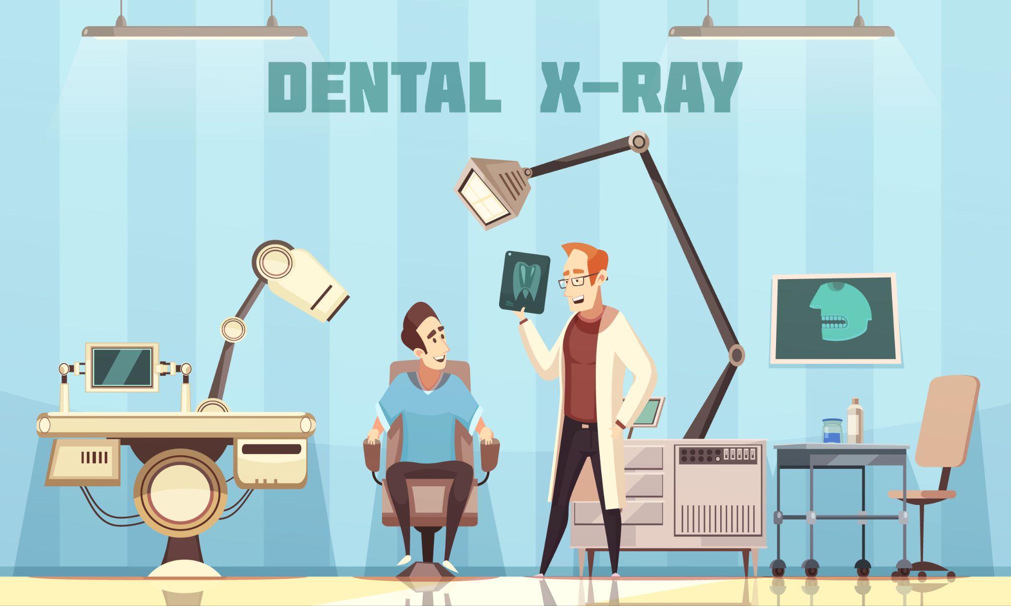
Dental X-rays or dental radiographs are images of your teeth and the jawbone. These images are used by dentists to examine areas of the mouth they can’t see during a routine checkup, like the teeth roots, jawbone, nerves, and even sinuses.
How Do Dental X-Rays Work?
Dental X-rays use electromagnetic waves to generate images of your mouth like any other X-rays. The X-ray machine passes a radiation beam, creating images of your teeth and jawbone.
There are two ways to generate a dental X-ray image – traditional and digital. Traditional X-rays are produced using film, and digital X-rays are created using digital sensors and computer imaging technology. Digital radiography was introduced in the mid‐1980s and operates with over 80% less radiation than traditional ones.

Types Of Dental X-Rays
Let’s look at the different types of dental X-rays, their purposes, and when you might need them.
Bitewing X-Ray
Bitewing X-rays are the most frequently used dental X-rays, usually taken for preventive care. They allow a closer look at any tooth decay or gum problems. For this, patients bite down on the X-ray film, which is then developed, hence the name, bitewing X-ray. This process can be done entirely on the dental chair.
Bitewing X-rays are also helpful in locating the source of toothache. Many modern dental clinics have switched to special sensors, which send dental X-rays directly to the computer for review by the dentist. This allows for quicker processing as the film doesn’t have to be developed.
Occlusal X-Ray
Occlusal X-rays provide a broader picture of everything that goes inside the roof and floor of the mouth, helping the dentist learn about the development and placement of the tooth. These insights are used to determine the reason behind unerupted teeth or spot any potential extra teeth, which can be bad for healthy permanent teeth.
To perform this X-ray, an occlusal radiograph film is placed between your upper and lower jaw and the dental X-ray tube head is positioned above the patient at an angle toward the X-ray film. Occlusal X-ray can also diagnose a fracture, cleft palate, cysts, and other potentially harmful growths.
Periapical X-Ray
Periapical X-rays are a type of dental X-ray done when a dentist needs to look at an entire tooth or jawbone. This type of X-ray captures an image of the whole tooth, from the crown to a little past the root tip.
Periapical radiography typically produces the image of the upper or lower row of teeth on a single film. This helps your dentist detect tooth decay, gum disease, root damage, bone loss, and other anomalies in your teeth or jawbone.
Panoramic X-Ray
As the name suggests, a panoramic X-ray produces one single image of your teeth. It is also known as an OPG radiograph. Radiographers use a particular machine that takes a 2-D image of your mouth. Panoramic dental X-rays are recommended when a patient experiences recurring dental complications or has had major dental work (like surgery) done in the past.
Getting a panoramic X-ray regularly helps get insights about any problems that may arise. Apart from that, dentists also use it when preparing for major dental procedures like getting braces. Serious issues such as jaw tumours, cysts, and sinusitis can also be determined using panoramic dental X-rays.
Cone Beam Computed Tomography (CBCT)
Cone beam dental X-rays are an advanced radiography method that uses computerised technology to convert two-dimensional (2D) images into three-dimensional (3D) pictures. The 3D print allows a view of every aspect of the teeth and the surrounding jawbone. Cone-beam X-ray is used to investigate the exact location of dental problems such as tumours and injuries. It also examines the impacted teeth before oral and maxillofacial surgery.
Compared to a 2D X-ray that shows a flat image, CBCT combines all areas of the mouth to produce a consolidated image. This saves examination time as you don’t have to sit for separate dental X-rays for each area of your mouth.
Cephalometric Projection
When a dentist takes an X-ray of one side of the patient’s entire head, it is called cephalometric projection. This type of dental X-ray is common with orthodontists to examine how the teeth and jawbones fit. This helps the dentist devise a treatment plan that covers the entire mouth.
A cephalometric projection is not just used for dental complications; it is also helpful in diagnosing severe throat problems like lumps or cancer. It can also be used to determine the cause of sleep apnea.
Are you due for a dental checkup? Dentists recommend regular dental cleanings and examinations to maintain optimal oral health. However, dental checkups are not just about cleaning; they also include using advanced technology to address dental problems. If you are looking for a dental clinic in Delhi, India, Kamal Dental Clinic is a trustworthy partner for all your dental needs. We use cutting-edge dental technology to provide safe and effective treatments.